
小鼠 单克隆 (PC 10)
反应物种: 人类, 小鼠, 大鼠
应用: 免疫印迹, 免疫组化, 免疫细胞化学
反应物种: 人类, 小鼠, 大鼠
应用: 免疫印迹, 免疫组化, 免疫细胞化学


Figure 1. Western blot analysis of PCNA using anti- PCNA antibody (MA1083). Electrophoresis was performed on a 5-20% SDS-PAGE gel at 70V (Stacking gel) / 90V (Resolving gel) for 2-3 hours. The sample well of each lane was loaded with 50ug of sample under reducing conditions. Lane 1: human Caco-2 whole cell lysates,. Lane 2: human MDA-MB-231 whole cell lysates, . Lane 3: human Jurkat whole cell lysates, . Lane 4: human HT1080 whole cell lysates. After Electrophoresis, proteins were transferred to a Nitrocellulose membrane at 150mA for 50-90 minutes. Blocked the membrane with 5% Non-fat Milk/ TBS for 1.5 hour at RT. The membrane was incubated with mouse anti- PCNA antigen affinity purified monoclonal antibody (Catalog # MA1083) at 0.5 ug/mL overnight at 4°C, then washed with TBS-0.1%Tween 3 times with 5 minutes each and probed with a goat anti- mouse IgG-HRP secondary antibody at a dilution of 1:10000 for 1.5 hour at RT. The signal is developed using an Enhanced Chemiluminescent detection (ECL) kit (Catalog # EK1001) with Tanon 5200 system. A specific band was detected for PCNA at approximately 35KD. The expected band size for PCNA is at 29KD.

Figure 2. IHC analysis of PCNA using anti- PCNA antibody (MA1083). PCNA was detected in paraffin-embedded section of human Rectal cancer tissues. Heat mediated antigen retrieval was performed in citrate buffer (pH6, epitope retrieval solution) for 20 mins. The tissue section was blocked with 10% goat serum. The tissue section was then incubated with 1ug/ml mouse anti- PCNA Antibody (MA1083) overnight at 4°C. Biotinylated goat anti-mouse IgG was used as secondary antibody and incubated for 30 minutes at 37°C. The tissue section was developed using Strepavidin-Biotin-Complex (SABC)(Catalog # SA1021) with DAB as the chromogen.

Figure 3. IHC analysis of PCNA using anti- PCNA antibody (MA1083). PCNA was detected in immunocytochemical section of human HELA Cell. Heat mediated antigen retrieval was performed in citrate buffer (pH6, epitope retrieval solution) for 20 mins. The tissue section was blocked with 10% goat serum. The tissue section was then incubated with 1ug/ml mouse anti- PCNA Antibody (MA1083) overnight at 4°C. Biotinylated goat anti- mouse IgG was used as secondary antibody and incubated for 30 minutes at 37°C. The tissue section was developed using Strepavidin-Biotin-Complex (SABC)(Catalog # SA1021) with DAB as the chromogen.
规格: 100μg/vial
价格: 315美元
至产商
domestic rabbit 单克隆 (DO-16)
反应物种: 人类, 小鼠, 大鼠
应用: 免疫印迹, 免疫组化, 免疫细胞化学, 免疫沉淀, 流式细胞仪
反应物种: 人类, 小鼠, 大鼠
应用: 免疫印迹, 免疫组化, 免疫细胞化学, 免疫沉淀, 流式细胞仪

Western blot analysis of PCNA expression in (1) Hela cell lysate; (2) HepG2 whole cell lysate; (3) U937 whole cell lysate; (4) Mouse spleen lysate with PCNA Antibody (M00125-1). Electrophoresis was performed on a 5-20% SDS-PAGE gel at 70V (Stacking gel) / 90V (Resolving gel) for 2-3 hours. The sample well of each lane was loaded with 50ug of sample under reducing conditions. After Electrophoresis, proteins were transferred to a Nitrocellulose membrane at 150mA for 50-90 minutes. Blocked the membrane with 5% Non-fat Milk/ TBS for 1.5 hour at RT. The membrane was incubated with rabbit anti-PCNA monoclonal antibody (Catalog # M00125-1) overnight at 4°C, then washed with TBS-0.1%Tween 3 times with 5 minutes each and probed with a goat anti-rabbit IgG-HRP secondary antibody at a dilution of 1:10000 for 1.5 hour at RT. The signal is developed using an Enhanced Chemiluminescent detection (ECL) kit (Catalog # EK1002) with Tanon 5200 system. A specific band was detected for PCNA

Immunohistochemical analysis of paraffin-embedded human colon, using PCNA Antibody(M00125-1). PCNA was detected in paraffin-embedded tissue section. Heat mediated antigen retrieval was performed in citrate buffer (pH6, epitope retrieval solution) for 20 mins. The tissue section was blocked with 10% goat serum. The tissue section was then incubated with 1ug/ml rabbit anti-PCNA Antibody (M00125-1)overnight at 4°C. Biotinylated goat anti-rabbit IgG was used as secondary antibody and incubated for 30 minutes at 37°C. The tissue section was developed using Strepavidin-Biotin-Complex (SABC)(Catalog # SA1022) with DAB as the chromogen.
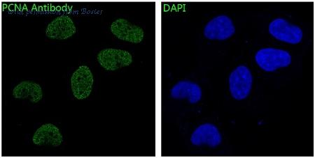
IF analysis of immunocytochemical section of Hela cells using anti- PCNA antibody (M00125-1) . PCNA was detected in immunocytochemical section. Enzyme antigen retrieval was performed using IHC enzyme antigen retrieval reagent (AR0022) for 15 mins. The tissue section was blocked with 10% goat serum. The tissue section was then incubated with 2ug/mL rabbit anti- PCNA Antibody (M00125-1) overnight at 4 C. DyLight®488 Conjugated Goat AntiRabbit IgG (BA1127) was used as secondary antibody at 1:100 dilution and incubated for 30 minutes at 37 C. The section was counterstained with DAPI. Visualize using a fluorescence microscope and filter sets appropriate for the label used.
规格: 100微升
价格: 315美元
至产商
domestic rabbit 多克隆
反应物种: 人类, 小鼠, 大鼠
应用: 免疫印迹, 酶联免疫吸附测定, 流式细胞仪, 免疫组化-石蜡切片
反应物种: 人类, 小鼠, 大鼠
应用: 免疫印迹, 酶联免疫吸附测定, 流式细胞仪, 免疫组化-石蜡切片
小鼠 单克隆 (2G2)
反应物种: 人类
应用: 免疫印迹, 免疫细胞化学, 流式细胞仪, 免疫组化-石蜡切片
反应物种: 人类
应用: 免疫印迹, 免疫细胞化学, 流式细胞仪, 免疫组化-石蜡切片
微博分享
关注我们的微博
- 来邦网
- 英文来邦